Total Pageviews

Wednesday 21 September 2011

Testing for H-pylori infection


diasource-diagnostics.com
The common tests are:


1.Serological testing
2.Urea breath test
3.Biopsy based urease
4.Histology
5.Microbial culture


1/ This detects antibody (IgG) response to H.pylori antigens. This confirms exposure at some point but will not discriminate between current or previous exposure. Therefore it can not be used to follow up eradication therapy.
2/This relies on bacterial urease activity in the stomach which is only found if H.pylori is present. Radiolabelled urea is ingested and is then broken down by the urease. Radiolabelled carbon dioxide is released into the blood and subsequently exhaled.  This test can be used to asses active infection but has time and cost implications in primary care.
3/This uses the same principle as the urea breath test. Tissue biopsy containing urease is placed in a urea solution. Ammonia is released which raises the pH of the solution which is detected by an indicator. This is the basis of the CLO (campylobacter like organism) test. A major diadvantage of this test is that it involves an invasive endoscopic procedure.
4/This requires visualisation of H.pylori adherent to the gastric mucosa. The bacteria can be directly viewed using HandE staining. Alternatively, specific stains or immunological techniques can be used. Disadvantage: the test is invasive. Advantage:  culture and sensitivity to the most commonly used agents (clarithromycin and metronidazole) can be performed.
5/Faecal antigen testing requires the detection of H.pylori antigens shed from live bacteria into the gut lumen.
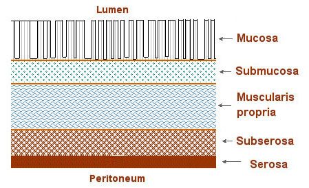
Duke's staging of colorectal cancer


training.seer.cancer.gov

A: Tumour which does not extend beyond muscularis propria (layer of circular and longitudinal muscles) à 97% five year survival

B: Tumour extends across muscularis propria with no nodal involvement à 80% five year survival but dramatically worse prognosis if locally invasive

C1: Full thickness growth. Only a few nodes are involved near the primary growth  and the proximal nodes are free from metastasis à 60% 5 year survival

C2: Proximal nodes are involved à 30% five year survival

D: Distant metastasis à <5% 5 year survival

Friday 16 September 2011

The 4 classical presentations of glomerulonephritis.

This is a notoriously complicated topic. However, a basic appreciation of the major presentations of glomerulonephritis should suffice for final exams.


1-Acute nephritic syndrome
2-Asymptomatic haematuria and / or proteinuria
3-Nephrotic syndrome
4-Chronic renal failure

1/ The acute nephritic syndrome is manifest by haematuria, hypertension, oliguria, proteinuria and sometimes oedema. This is most often seen with either acute proliferative glomerulonephritis (often post streptococcal) or diffuse proliferative crescenteric glomerulonephritis (seen in systemic vasculitis and rarely anti GBM disease).

2/This is typical of IgA nephropathy when recurrent heavy haematuria can occur following URTI. Asymptomatic proteinuria can also be seen with IgA nephropathy or the membranoproliferative glomerulonephritides.

3/ Characterised by heavy proteinuria (>3.5g day), hypoalbuminaemia, oedema and hypercholesterolaemia. This is most commonly associated with minimal change disease in children and membranous glomerulonephritis in adults.

4/  Chronic renal failure from glomerulonephritis may follow from symptomatic acute nephritic syndrome eg due to systemic vasculitis with crescenteric glomerulonephritis or it may follow on from the recurrent haematuria of IgA nephropathy. Chronic renal failure with significant elevations of urea and creatinine may also be diagnosed de novo and be due to the progression of preexisting glomerulonephritis. In some cases a diagnosis cannot be made because the end stage atrophic kidneys preclude diagnosis.


glufkids.com
A child with nephrotic syndrome